Past Projects
PAST 3-D CELL/TISSUE ORGANOID-LIKE PROJECTS
Cell Systems 3-D has designed and bioengineered numerous prototype 3-D models for investigations in the pathophysiology of microbial, viral and fungi pathogens.
These 3-D models have been used in NIH or privately sponsored research.
VIROLOGY
- Proof of concept; human cerebellum 3-D model for invasiveness and pathology of West Nile Virus, (NY-99 strain) and Dengue-2.
- Pathophysiology of SARS-coronavirus (SARS-CoV, Urbani strain) in a human upper airways 3-D model.
- Pathophysiology of Respiratory Syncytial Virus (wt-A2) and proof of concept for Parainfluenza 1 and 3 in a 3-D human airways model.
- Pathophysiology of Epstein-Barr Virus (EBV) in a human endovascular – pharyngeal epithelia 3-D model.
- Proof of concept 3-D mosquito Ae. albopictus 3-D model for arbovirus transmission.
MICROBIAL
- Proof of concept; 3-D human colon model for Mycobacterium spp. invasiveness and pathophysiology.
PARASITOLOGY
- Proof of concept; normal human dermis providing a microenvironment for filarid nematode development from infective larvae to adult.
- Proof of concept; Human small intestine model for life cycle stage development of Cryptosporidium parvum.
VETERINARY
- Proof of concept; avian chick small intestinal model for selected bacterial pathogens.
- Proof of concept; neonatal bovine small intestine-colon for selected bacterial pathogens.
- Proof of concept; tick mid-gut cell model for proteomic comparisons between in-stage and adult tissue.
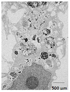
Human upper airways 3-D model. Bronchial epithelial cell infected with SARS-CoV, day 4 post-infection.
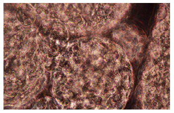
Three-dimensional (3-D) tick mid-gut intestinal cells adhering to macrogelantinous beads. x400